
Sign In
Conformal Ultrasound Patch for Accurate and Continuous Transcranial Monitoring of Cerebral Blood Flow
Core Concepts
A conformal ultrasound patch enables accurate and continuous volumetric imaging and monitoring of cerebral blood flow, overcoming the limitations of conventional transcranial Doppler ultrasonography.
Abstract
The content describes the development and evaluation of a conformal ultrasound patch for transcranial volumetric imaging and continuous monitoring of cerebral blood flow. Key highlights:
Conventional transcranial Doppler (TCD) ultrasonography has limitations in accurately measuring the complex 3D vascular networks and practicality for prolonged recording.
The conformal ultrasound patch uses 2 MHz ultrasound waves to reduce attenuation and phase aberration caused by the skull, and a copper mesh shielding layer to improve signal-to-noise ratio.
Ultrafast ultrasound imaging based on diverging waves can accurately render the circle of Willis in 3D and minimize human errors during examinations.
Focused ultrasound waves allow continuous recording of blood flow spectra at selected locations.
Evaluation on 36 participants showed the conformal patch had a higher measurement success rate (70.6%) compared to a conventional TCD probe (75.3%), with high accuracy in measuring peak systolic velocity, mean flow velocity, and end diastolic velocity.
The patch enabled continuous monitoring of blood flow spectra during different interventions and identification of intracranial B waves during drowsiness.
Transcranial volumetric imaging using a conformal ultrasound patch - Nature
Stats
The conformal ultrasound patch showed a mean difference and standard deviation of difference compared to a conventional TCD probe of:
-1.51 ± 4.34 cm/s for peak systolic velocity
-0.84 ± 3.06 cm/s for mean flow velocity
-0.50 ± 2.55 cm/s for end diastolic velocity
Quotes
"Accurate and continuous monitoring of cerebral blood flow is valuable for clinical neurocritical care and fundamental neurovascular research."
"The high accuracy of the conformal ultrasound patch was confirmed in comparison with a conventional TCD probe on 36 participants."
"Furthermore, we demonstrate continuous blood flow spectra during different interventions and identify cascades of intracranial B waves during drowsiness within 4 h of recording."
Key Insights Distilled From
by Sai Zhou,Xia... at www.nature.com 05-22-2024
https://www.nature.com/articles/s41586-024-07381-5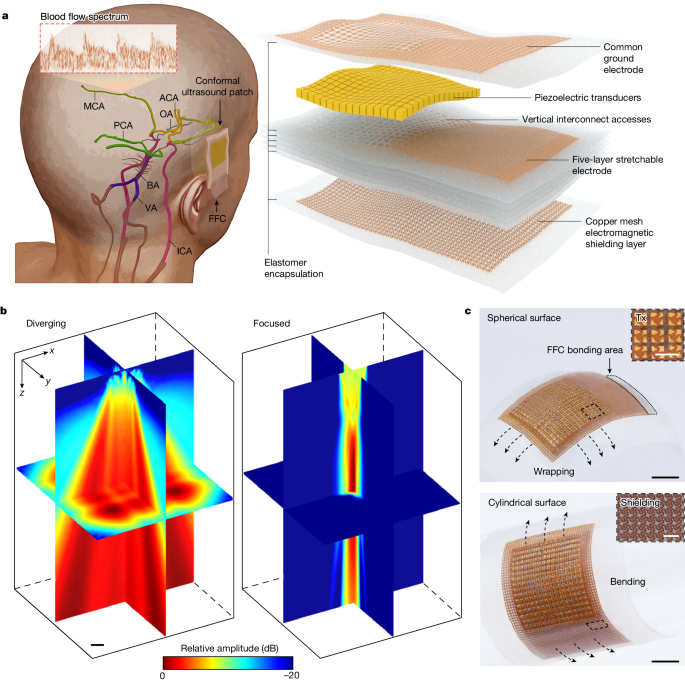
Deeper Inquiries
How can the conformal ultrasound patch be further improved to increase the measurement success rate and expand its clinical applications?
To enhance the conformal ultrasound patch's performance and applicability, several improvements can be considered. Firstly, optimizing the ultrasound frequency range could improve penetration depth and resolution, enabling better visualization of deeper cerebral structures. Additionally, incorporating real-time image processing algorithms to automatically detect and track blood flow patterns could reduce human error and enhance measurement accuracy. Moreover, integrating wireless connectivity for data transmission would enhance the patch's usability in various clinical settings, allowing for remote monitoring and analysis. Furthermore, exploring the integration of additional sensors, such as temperature or pressure sensors, could provide complementary physiological data for a more comprehensive assessment of cerebral hemodynamics.
What are the potential limitations or drawbacks of using a conformal ultrasound patch compared to other neuroimaging techniques for cerebral blood flow monitoring?
While the conformal ultrasound patch offers significant advantages, such as non-invasiveness and continuous monitoring capabilities, it also has limitations compared to other neuroimaging techniques. One potential drawback is the limited depth penetration of ultrasound waves, which may restrict visualization of deep brain structures or small vessels. Additionally, the reliance on acoustic windows for optimal imaging quality could pose challenges in patients with anatomical variations or skull defects. Moreover, the need for direct skin contact with the patch may limit its application in patients with skin conditions or wounds. Furthermore, the current technology may have limitations in differentiating between arterial and venous blood flow, which could impact the accuracy of cerebral blood flow measurements in certain clinical scenarios.
What other physiological or neurological insights could be gained by continuously monitoring cerebral blood flow patterns using the conformal ultrasound patch technology?
Continuous monitoring of cerebral blood flow patterns using the conformal ultrasound patch technology could provide valuable insights into various physiological and neurological processes. By analyzing dynamic changes in blood flow velocities, researchers and clinicians can gain a deeper understanding of cerebral autoregulation mechanisms and vascular reactivity in response to different stimuli. Furthermore, continuous monitoring could help identify subtle hemodynamic alterations associated with conditions such as vasospasm, ischemia, or hyperperfusion, enabling early detection and intervention. Additionally, studying the temporal patterns of blood flow spectra could offer insights into neurovascular coupling mechanisms and the impact of cerebral perfusion on cognitive function and brain metabolism. Overall, continuous monitoring of cerebral blood flow using the conformal ultrasound patch has the potential to uncover novel biomarkers and physiological relationships relevant to neurological health and disease.
0
More on Medical Imaging
Products | Resources
Read more
- comprehensive-evaluation-of-few-shot-learning-techniques-and-pre-trained-language-models-for-polish-classification-tasks
- ポーランド語の分類タスクにおけるフューショットラーニングの評価
- 폴란드어-분류-작업을-위한-소량-학습-평가
- automated-testing-and-analysis-of-erroneous-planning-in-large-language-model-agents-through-synthesized-user-inputs
- llm代理的错误规划检测和理解-基于合成用户输入
- llm-에이전트의-잘못된-계획을-합성된-사용자-입력을-통해-테스트하고-이해하기
- polynomial-time-algorithm-for-realizing-expressive-temporal-logic-specifications
- 生産ラインの効率的な制御合成
© 2024 by Linnk AI