
Sign In
Imaging Techniques Demonstrate Comparable Accuracy to Dilated Fundus Exams for Detecting Retinal Tears in Patients with Acute Posterior Vitreous Detachment
Core Concepts
Alternative imaging techniques, including fundus photography and ultrasonography, can accurately identify retinal tears in patients with acute posterior vitreous detachment, potentially enabling easier access to care and telemedicine-based diagnosis.
Abstract
The study presented at the 2024 ARVO annual meeting evaluated the accuracy of fundus photography and 20-MHz B-scan ultrasonography in detecting retinal tears in 35 eyes of patients with acute posterior vitreous detachment (aPVD). The researchers found that both techniques showed "non-inferiority" to the current gold standard of dilated fundus exams with indentation by retina specialists.
The B-scan ultrasonography had a sensitivity of 100% and a specificity of 96.6% in detecting retinal tears, while fundus photography had a sensitivity of 83.3% and a specificity of 100%. Optical coherence tomography (OCT) was also accurate in confirming the diagnosis of posterior vitreous detachment.
The researchers noted that the use of these alternative imaging techniques could enable easier access to care, including through telemedicine, for patients with aPVD who may not have immediate access to ophthalmology clinics. While the current sample size was small, the researchers plan to expand the study to 250 participants to further validate the findings.
Imaging Techniques May Match Gold Standard for Retinal Tears
Stats
17% of the 35 eyes examined had retinal tears.
B-scan ultrasonography had a sensitivity of 100% and a specificity of 96.6% in detecting retinal tears.
Fundus photography had a sensitivity of 83.3% and a specificity of 100% in detecting retinal tears.
Quotes
"Even though the sample size was small, the results are promising and open the grounds for using telemedicine for those who do not have access to ophthalmology clinics and to provide timely referral for treatment."
"While they could be used separately, they are even more accurate when used in combination."
Key Insights Distilled From
by Jake Remaly at www.medscape.com 05-15-2024
https://www.medscape.com/viewarticle/imaging-techniques-may-match-gold-standard-retinal-tears-2024a10009as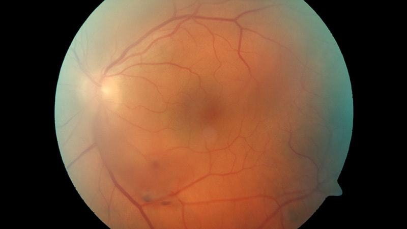
Deeper Inquiries
How could the integration of these alternative imaging techniques into telemedicine platforms improve access to timely diagnosis and treatment of retinal tears for patients in underserved or remote areas?
The integration of alternative imaging techniques such as fundus photography and B-scan into telemedicine platforms can significantly enhance access to timely diagnosis and treatment of retinal tears for patients in underserved or remote areas. By utilizing these imaging modalities, healthcare providers can remotely assess and accurately identify retinal tears without the need for in-person visits to ophthalmology clinics. This is particularly beneficial for individuals in areas with limited access to specialized eye care services. Telemedicine allows for the rapid transmission of images to ophthalmologists for review, enabling prompt diagnosis and referral for treatment. This approach can expedite the initiation of appropriate interventions, ultimately improving patient outcomes and reducing the risk of complications associated with untreated retinal tears.
What potential limitations or challenges might arise in the widespread adoption of these imaging techniques, and how could they be addressed?
Despite their potential benefits, the widespread adoption of alternative imaging techniques for diagnosing retinal tears may face certain limitations and challenges. One key challenge is the availability and accessibility of the necessary equipment, such as the 20-MHz B-scan annular array used in the study. This specialized equipment may not be widely accessible in all healthcare settings, particularly in underserved areas. Additionally, the requirement for technical training to perform B-scan imaging and the variability in results based on the operator's expertise pose potential challenges to widespread adoption. To address these limitations, efforts should be made to increase the availability of imaging equipment in healthcare facilities, provide training programs for healthcare professionals on using the technology effectively, and ensure quality control measures to maintain the accuracy and reliability of imaging results. Collaborative initiatives involving healthcare institutions, government agencies, and technology providers can help overcome these challenges and facilitate the broader implementation of these imaging techniques.
Given the differences in sensitivity and specificity between the techniques, how could clinicians best utilize a multimodal approach to optimize the accuracy of retinal tear diagnosis?
Clinicians can optimize the accuracy of retinal tear diagnosis by strategically utilizing a multimodal approach that combines different imaging techniques, taking into account their respective strengths and limitations. In the context of detecting retinal tears, the study found that B-scan imaging demonstrated high sensitivity and specificity, making it a valuable screening tool. On the other hand, fundus photography showed high specificity but slightly lower sensitivity, making it more suitable as a confirmatory test. By integrating these modalities, clinicians can leverage the strengths of each technique to complement and validate the findings of the other. For instance, clinicians can use B-scan as an initial screening tool to identify potential retinal tears, followed by fundus photography to confirm the diagnosis with higher specificity. This sequential approach can enhance diagnostic accuracy and reduce the likelihood of false-positive or false-negative results. Additionally, incorporating optical coherence tomography (OCT) for confirming posterior vitreous detachment (PVD) can further enhance the diagnostic precision of the multimodal approach. By judiciously combining these imaging modalities, clinicians can optimize the accuracy of retinal tear diagnosis and provide more comprehensive care to patients.
0
Visualize This Page
Generate with Undetectable AI
Translate to Another Language
Scholar Search
Products | Resources
© 2024 by Linnk AI