
Sign In
Core Concepts
PET scans at GCA diagnosis can predict aneurysm risk.
Abstract
In a study focusing on giant cell arteritis (GCA), PET scans were utilized as both a diagnostic and prognostic tool. The study followed over 100 GCA patients who underwent 18F-fluorodeoxyglucose (FDG) PET imaging, revealing that those with elevated FDG uptake at diagnosis were more prone to developing thoracic aortic aneurysms. The research, led by Lien Moreel, MD, from the University Hospitals Leuven, Belgium, emphasizes the potential of PET imaging at diagnosis to estimate the future risk of aortic aneurysm formation in GCA patients. The study, published in Annals of Internal Medicine, highlights the importance of early PET scans for diagnostic and prognostic purposes in GCA patients. The positive PET scan results were associated with a higher risk of incident thoracic aortic aneurysms, indicating the predictive value of PET imaging in assessing aortic complications in GCA patients. The study suggests the need for further research to establish clear guidelines for the follow-up and monitoring of GCA patients with positive PET scan results.
PET Scan at Diagnosis May Help Predict Aneurysm Risk in GCA
Stats
"In the study, Moreel and colleagues prospectively followed 106 individuals diagnosed with GCA who received FDG-PET within 3 days after starting glucocorticoids."
"A PET scan was considered positive with an FDG uptake of grade 2 or higher in any of seven vascular regions."
"The risk of incident thoracic aortic aneurysms was calculated to be 10 times higher in patients with positive PET scans."
Quotes
"PET-CT has an excellent diagnostic accuracy for the diagnosis of GCA, certainly if both extracranial and intracranial vessels were assessed." - Lien Moreel, MD
"There are no clear guidelines, but most clinicians who take care of patients with GCA do obtain imaging periodically." - Dr. Kenneth Warrington
Key Insights Distilled From
by Lucy Hicks at www.medscape.com 10-02-2023
https://www.medscape.com/viewarticle/997028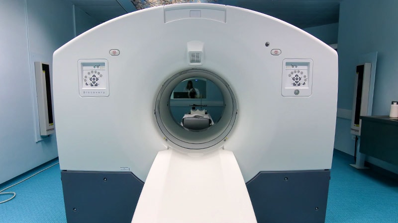
Deeper Inquiries
How can the findings of this study impact the current diagnostic and monitoring practices for GCA patients?
The findings of this study can significantly impact the current diagnostic and monitoring practices for patients with Giant Cell Arteritis (GCA). By utilizing PET scans at diagnosis, clinicians can not only accurately diagnose GCA but also predict the future risk of aortic aneurysm formation in these patients. This dual functionality of PET imaging provides valuable prognostic information that can guide treatment decisions and monitoring strategies. Specifically, patients with elevated FDG uptake on PET scans at diagnosis were more likely to develop thoracic aortic aneurysms, indicating the potential for early identification of high-risk individuals. This information can lead to more targeted and proactive management of GCA patients, potentially reducing the incidence of serious complications such as aortic aneurysms.
What are the potential limitations or challenges in implementing routine PET scans for GCA patients in clinical settings?
While the use of PET scans in GCA patients offers valuable diagnostic and prognostic information, there are several limitations and challenges to consider in implementing routine PET scans in clinical settings. One major limitation is the cost associated with PET imaging, which can be a barrier for widespread adoption, especially in healthcare systems with limited resources. Additionally, logistical challenges such as the need to perform imaging within a specific timeframe (e.g., within 3 days of starting glucocorticoids) can be difficult to meet in practice. Furthermore, insurance coverage for PET scans may vary, posing challenges for patients in accessing this imaging modality. These limitations highlight the need for alternative approaches or strategies to overcome barriers to routine PET imaging in GCA patients.
How might the use of alternative imaging modalities influence the predictive capabilities of assessing aortic complications in GCA patients?
The use of alternative imaging modalities, such as CT or MRI, in assessing aortic complications in GCA patients can provide valuable insights into the predictive capabilities of identifying at-risk individuals. While PET scans offer high sensitivity and specificity in detecting vascular inflammation, the feasibility and accessibility of alternative imaging modalities like CT or MRI may make them more practical in certain clinical settings. By extrapolating findings from PET scans to these modalities, clinicians can still assess the presence of aortic inflammation and potentially predict the risk of aortic complications in GCA patients. Although the specificity and accuracy of alternative imaging modalities may vary compared to PET scans, their use can still contribute to the early identification and monitoring of aortic complications in GCA patients, thereby enhancing the overall management of the disease.
0
Visualize This Page
Generate with Undetectable AI
Translate to Another Language
Scholar Search
Products | Resources
Read more
- 고속-분류-및-회귀-객체-클래스의-연속-속성-변수-모델링을-위한-다중-작업-학습
- 문장-기반-4d-생성을-위한-궤적-조건화
- 효율적인-트랜스포머-모델-압축을-위한-one-shot-프루닝-기법
- schnelle-klassifizierung-und-regression-ein-neuartiger-ansatz-zur-aufgabenkonsolidierung-im-multi-task-lernen-für-die-modellierung-kontinuierlicher-objekteigenschaften
- effiziente-generierung-von-4d-szenen-mit-globaler-und-lokaler-bewegung-durch-trajektorien-konditionierung
- effiziente-kompression-von-transformer-architekturen-durch-one-shot-pruning
- llmを活用したgithubイシューの解決のための多エージェントフレームワーク-magis-
- ハイパーコンプレックスニューラルネットワークの解釈可能性を高める
© 2024 by Linnk AI