
Detailed 3D Microscopy Reveals the Intricate Organization of Amyloid-β Plaques and Tau Tangles in Alzheimer's Disease
The content discusses how the hallmark pathological features of Alzheimer's disease, namely amyloid-β plaques and tau tangles, are organized at the molecular level. Previous light microscopy studies have revealed the presence of these protein aggregates, but the precise arrangement of individual filaments within the plaques and tangles has remained unclear.
The authors used a technique called cryo-electron tomography to obtain high-resolution 3D images of brain tissue from Alzheimer's patients. This allowed them to visualize the intricate organization of amyloid-β and tau filaments that make up the plaques and tangles, respectively.
Key insights from the 3D microscopy data:
- Amyloid-β filaments assemble into a highly organized, fibrillar structure within the plaques.
- Tau filaments form twisted, intertwined strands that bundle together to create the characteristic tangles inside brain cells.
- The spatial arrangement of these filamentous assemblies provides important clues about the pathogenic mechanisms underlying Alzheimer's disease.
Overall, this study leverages cutting-edge imaging technology to shed unprecedented light on the molecular architecture of the hallmark Alzheimer's pathologies, which could inform the development of targeted therapies.
Customize Summary
Rewrite with AI
Generate Citations
Translate Source
To Another Language
Generate MindMap
from source content
Visit Source
www.nature.com
Alzheimer’s plaques and tangles revealed by 3D microscopy
Key Insights Distilled From
by Sjors H. W. ... at www.nature.com 07-10-2024
https://www.nature.com/articles/d41586-024-02119-9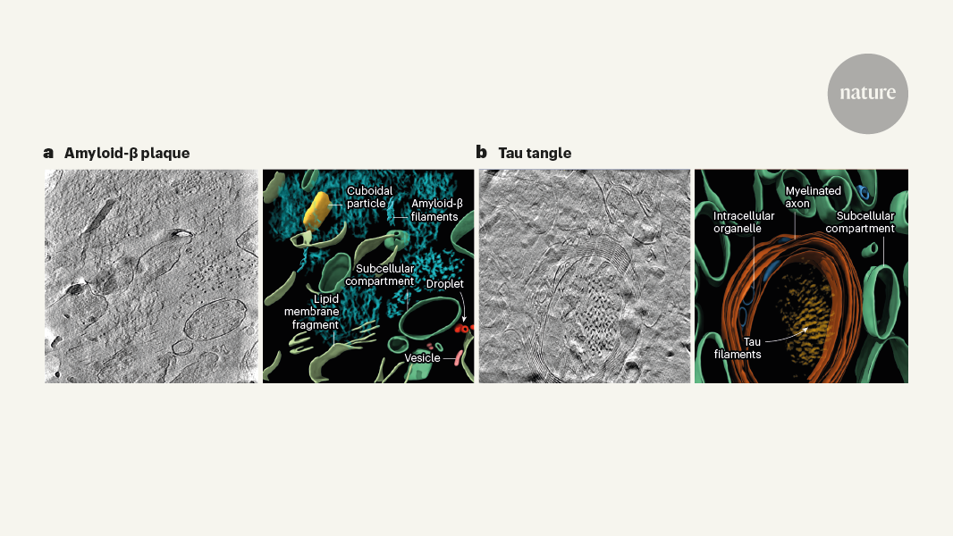
Deeper Inquiries
How do the structural features of amyloid-β plaques and tau tangles revealed by cryo-electron tomography relate to their proposed mechanisms of toxicity and neurodegeneration?
What are the potential limitations or caveats of the cryo-electron tomography approach used in this study, and how might they be addressed in future research?
Could the insights gained from visualizing Alzheimer's pathology at the molecular scale inspire the development of novel therapeutic strategies targeting the underlying protein aggregation processes?
© 2024 by Linnk AI